
Interrupted Aortic Arch (IAA)
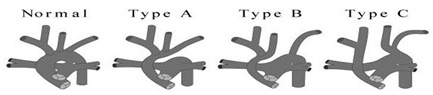
In interrupted aortic arch, there is an absence of luminal continuity between the ascending aorta and the descending aorta. There are three types of IAA, and each describes where the interruption takes place. Most common type is B, where the defect lies between the left carotid and left subclavian artery. There is almost always a large VSD. An aberrant right subclavian artery arising from the descending thoracic aorta and passing posterior to the esophagus is common, especially in type B. Blood reaches the descending aorta via the ductus arteriosus (PGE-1 dependent).
 Patients present within the first 2 weeks of life. As PVR decreases and pulmonary blood flow increases, patients develop signs of congestive heart failure, and these worsen as the ductus begins to close. The lower extremities may also become mottled or gray. There is eventually circulatory collapse and profound shock. PGE1 should be infused to maintain ductal patency. Hyperventilation should be avoided so as to not increase pulmonary blood flow and further worsen systemic perfusion. Echocardiography is used for diagnosis and can identify the site of the interruption as well as an anomalous right subclavian artery and any associated cardiac defects, including the VSD. DiGeorge syndrome is present about 27% of the time and may be indicated by hypocalcemia.
Repair is accomplished through a median sternotomy, and the arch may be reconstructed under deep hypothermic circulatory arrest, using a piece of homograft for augmentation if necessary. The PDA and the VSD are both closed in the same procedure.