Special Stains
There are a wide variety of special stains to demonstrate pathologic processes. They generally employ a dye or chemical which has an affinity for the particular tissue component to be demonstrated. Special Stains available include:
1. Connective Tissue Stains
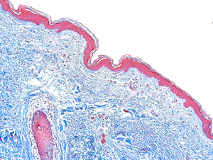
Massons Trichrome
The trichrome stain helps to highlight the supporting collagenous stroma in sections from a variety of organs. This helps to determine the pattern of tissue injury. Trichrome will also aid in identifying normal structures, such as connective tissue capsules of organs, the lamina propria of the gastrointestinal tract, and the bronchovascular structures in the lung.
Verhoff’s Elastic Stain
An elastic tissue stain helps to outline arteries, because the elastic lamina of muscular arteries, and the media of the aorta, contain elastic fibers. The van Gieson method for elastic fibers provides good contrast
Reticulin Stain
The reticulin stain is useful in parenchymal organs such as liver and spleen to outline the architecture. Delicate reticular fibers, which are argyrophilic, can be seen. A reticulin stain occasionally helps to highlight the growth pattern of neoplasms.
Giemsa Stain
There are a variety of “Romanowsky-type” stains with mixtures of methylene blue, azure, and eosin compounds. Among these are the giemsa stain and the Wright’s stain (or Wright-Giemsa stain). The latter is utilized to stain peripheral blood smears. The giemsa stain can be helpful for identifying components in a variety of tissues. One property of methylene blue and toluidine blue dyes is metachromasia. This means that a tissue component stains a different color than the dye itself. For example, mast cell graules, cartilage, mucin, and amyloid will stain purple and not blue, which is helpful in identifying these components.
2. Microrganisms
Brown and Hopps
Bacteria appear on H and E as blue rods or cocci regardless of gram reaction. Colonies appear as fuzzy blue clusters. Tissue gram stains are all basically the same as that used in the microbiology lab except that neutral red is used instead of safranin. Gram positive organisms usually stain well, but gram negatives do not (because the lipid of the bacterial walls is removed in tissue processing).
AFB (acid-fast bacilli) stain
This stain uses carbol-fuchsin to stain the lipid walls of acid fast organisms such as M. tuberculosis. The most commonly used method is the Ziehl-Neelsen method, though there is also a Kinyoun’s method. A modification of this stain is known as the Fite stain and has a weaker acid for supposedly more delicate M. leprae bacilli. However, much of the lipid in mycobacteria is removed by tissue processing, so this stain can, at times, be very frustrating and cause you to search extensively for organsisms you are sure are in a big granuloma. The most sensitive stain for mycobacteria is the auramine stain which requires a fluorescence microscope for viewing. There are things other than mycobacteria that are acid fast. Included are cryptosporidium, isospora, and the hooklets of cysticerci.
Gomori methenamine silver stain
This stain, often abbreviated as “GMS”, is used to stain for fungi and for Pneumocystis carinii. The cell walls of these organisms are stained, so the organisms are outlined by the brown to black stain. There is a tendency for this stain to produce a lot of artefact from background staining, so it is essential to be sure of the morphology of the organism being sought.
PAS (periodic acid-Schiff)
This an all-around useful stain for many things. It stains glycogen, mucin, mucoprotein, glycoprotein, as well as fungi. A predigestion step with amylase will remove staining for glycogen. PAS is useful for outlining tissue structures–basement membranes, capsules, blood vessels, etc. It does stain a lot of things and, therefore, can have a high background. It is very sensitive, but specificity depends upon interpretation.
3. Carbohydrate Stains
Colloidal iron (“AMP”)
Iron particles are stabilized in ammonia and glycerin and are attracted to acid mucopolysaccharides. It requires a formalin fixation. Phospholipids and free nucleic acids may also stain. The actual blue color comes from a Prussian blue reaction. Tissue can be pre-digested with hyaluronidase to provide more specificity.
Alcian blue
The pH of this stain can be adjusted to give more specificity.
PAS (peroidic acid-Schiff)
Stains glycogen as well as mucins, but tissue can be pre-digested with diastase to remove glycogen.
Mucicarmine
Very specific for epithelial mucins.
4. Pigments, Minerals and Cytoplasmic Granules
Fontana-Masson for Melanin
The method relies upon the melanin granules to reduce ammoniacal silver nitrate (but argentaffin, chromaffin, and some lipochrome pigments also will stain black as well).
Melanin Bleach
Bleaching techniques remove melanin in order to get a good look at cellular morphology. They make use of a strong oxidizing agent such as potassium permanganate or hydrogen peroxide. Ocular melanin takes hours to bleach, while that from skin takes minutes.
Iron Stain
The classic method for demonstrating iron in tissues. The section is treated with dilute hydrochloric acid to release ferric ions from binding proteins. These ions then react with potassium ferrocyanide to produce an insoluble blue compound (the Prussian blue reaction). Mercurial fixatives seem to do a better job of preserving iron in bone marrow than formalin.
VonKossa Stain
Silver reduction method that demonstrates phosphates and carbonates, but these are usually present along with calcium.
5. Fat Stains
Oil Red O (ORO)
Stain Identifies neutral lipids and fatty acids in smears and tissues. Fresh smears or cryostat sections of tissue are necessary because fixatives containing alcohols, or routine tissue processing with clearing, will remove lipids. ORO is a rapid and simple stain. It can be useful in identifying fat emboli in lung tissue or clot sections of peripheral blood.

